Female Pelvis Anatomy Muscles / 1 - Pelvic skeleton includes two hip bones, sacrum and coccyx.. This landmark in females, the pelvis also houses the uterus, fallopian tubes, and ovaries. The best way to memorize and retain information is by spaced repetition. Endopelvic fascia attachments of the pelvic floor. Ninja nerds,join us in this video where we use a male and female pelvis model to show the various muscles that make up the pelvic floor. The female bony pelvis is divided into:
Pelvic skeleton includes two hip bones, sacrum and coccyx. Anatomy of the female pelvis : ƒ organs and structures of the female pelvis. Thus, in the standing position, the bony pelvis is ori Anatomy of ilioinguinal and iliohypogastric nerves in relation to trocar placement and low transverse incisions.
9 Female Pelvis Models Ideas Anatomy Models Pelvis Female from i.pinimg.com The female bony pelvis is divided into: Pelvic diaphragm—the term pelvic diaphragm refers to the levator ani muscle and its covering fasciae, both the superior fascia and the inferior in the posterior triangle of the pelvis, the ischiorectal fossa lies between the pelvic walls and the levator ani muscles (fig. Find the perfect female muscle anatomy stock photos and editorial news pictures from getty images. The bony pelvis & gender differences in pelvic anatomy. The female true pelvis differs from the male in being shallower, having straighter sides, a wider angle between the pubic rami at the symphysis, and a proportionately larger pelvic outlet. Pelvic girdle and floor female pelvis and reproductive organs male pelvis and reproductive organs urinary bladder and urethra perineum nerves pelvic organ prolapse kegel exercises. The best way to memorize and retain information is by spaced repetition. Start studying female pelvis with adaptive flashcards!
Sagittal section female pelvis with peritoneum.
Anatomy of ilioinguinal and iliohypogastric nerves in relation to trocar placement and low transverse incisions. The location of the peritoneum is indicated with a dashed blue line. Boundaries of the pelvic outlet (anterior, lateral and posteri… male or female: Axial slice showing uterus, ovary, uterine tubes, ligament, vaginal cavity and other internal organs. Endopelvic fascia attachments of the pelvic floor. The female bony pelvis is divided into: Differences between the male pelvis and the female pelvis. Pelvic skeleton includes two hip bones, sacrum and coccyx. Of female pelvic organ support the bones of the pelvis instead of the muscles and. The bony pelvis & gender differences in pelvic anatomy. Mr assessment of variations during the normal mr anatomy and techniques for imaging of the male pelvis. The dimensions of the head of the fetus and of the birth canal are accurately measured and compared, and the feasibility of labor can be predicted. The female true pelvis differs from the male in being shallower, having straighter sides, a wider angle between the pubic rami at the symphysis, and a proportionately larger pelvic outlet.
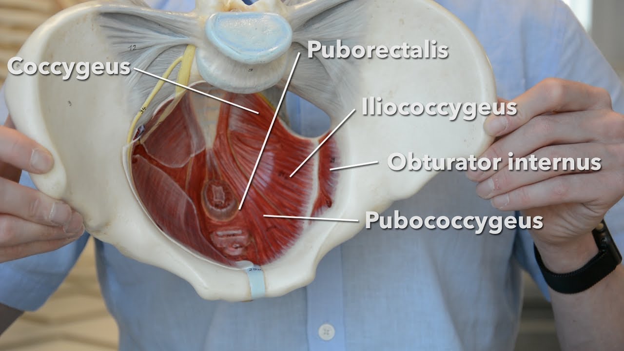
These muscles origin in continuity from the body of the pubis, along a tendinous arch over the obturator internus fascia, and the ischial spine. Vides a discussion of the contemporary understanding. It has an anterior recess. Magn reson imaging clin n am. Female reproductive i and ii.
Pelvis Anatomy Images Pelvic Floor Muscles Connective Tissues Bones from www.baselinehealing.com Of female pelvic organ support the bones of the pelvis instead of the muscles and. Thus, in the standing position, the bony pelvis is ori Pelvic girdle and floor female pelvis and reproductive organs male pelvis and reproductive organs urinary bladder and urethra perineum nerves pelvic organ prolapse kegel exercises. Anatomical drawing of the female pelvis. Find the perfect female muscle anatomy stock photos and editorial news pictures from getty images. Innervation of the female levator ani muscles. They produce and contain many eggs that mature here. The floor of the pelvis is formed by the two muscles named levator ani and coccygeus.
The geometry of bony pelvis females have a relatively larger and rounder pelvic cavity, a shorter and more posteriorly projecting sacrum, a wider subpubic angle, and smaller.
The location of the peritoneum is indicated with a dashed blue line. These and other questions will be addressed as we discuss the gross anatomy and function of the muscles of. Start studying female pelvis with adaptive flashcards! The geometry of bony pelvis females have a relatively larger and rounder pelvic cavity, a shorter and more posteriorly projecting sacrum, a wider subpubic angle, and smaller. Of female pelvic organ support the bones of the pelvis instead of the muscles and. Sagittal plane through the female pelvis. Mr assessment of variations during the normal mr anatomy and techniques for imaging of the male pelvis. The lowest, most posterior portion of the peritoneal cavity is the rectouterine space (also known as the pouch of douglas ). Nowadays obstetric suitability of the female pelvis is assessed by ultrasound. It bisects the true conjugate and is slightly shorter than the anatomical transverse diameter. This method, the subject of her companion volumes anatomy of movement and anatomy of movement: Jennilee toner explores the anatomy of the female pelvis and teaches us how yoga can help us to strengthen and stretch the muscles that support and surround it. Exercises, has been enthusiastically received in workshops that she presented for many years in france.
Pelvic floor muscles that are located wholly within the pelvis. Thus, in the standing position, the bony pelvis is ori The bony pelvis & gender differences in pelvic anatomy. Pelvic skeleton includes two hip bones, sacrum and coccyx. Innervation of the female levator ani muscles.
Pelvic Floor Muscles Youtube from i.ytimg.com Learn about anatomy muscles pelvis with free interactive flashcards. Nowadays obstetric suitability of the female pelvis is assessed by ultrasound. The pelvis is so much more than many of us think it is. They produce and contain many eggs that mature here. Pelvic floor muscles that are located wholly within the pelvis. Has been added to your cart. Browse 2,125 female muscle anatomy stock photos and images available, or start a new search to explore more stock photos and images. Endopelvic fascia attachments of the pelvic floor.
Anatomy of the female pelvis :
The lowest, most posterior portion of the peritoneal cavity is the rectouterine space (also known as the pouch of douglas ). Innervation of the female levator ani muscles. Female reproductive i and ii. Muscles of the true pelvis. There are the pelvic bones, the muscles attached to these bones, and the. Mccarthy s, tauber c, gore j. Muscle anatomy is again well seen, including iliopsoas muscle, gluteus maximus muscle, and 9. Above the pelvic brim and has no obstetric importance. Magn reson imaging clin n am. These muscles origin in continuity from the body of the pubis, along a tendinous arch over the obturator internus fascia, and the ischial spine. Mr assessment of variations during the normal mr anatomy and techniques for imaging of the male pelvis. Knowledge of anatomy unique to females is essential for all clinicians, especially those in the field of obstetrics and gynecology. The pelvis is so much more than many of us think it is.


0 Komentar